Purpose of This Research
Advancing diagnostic capabilities through AI and machine learning
Research Goal
Our research aims to develop accurate, accessible AI-based diagnostic tools for lung diseases, focusing on pneumonia and tuberculosis detection from chest X-rays. By leveraging machine learning technology, we strive to create solutions that can assist healthcare professionals in underserved regions where radiological expertise may be limited.
Clinical Significance
Lung diseases like pneumonia and tuberculosis affect millions globally, with early detection crucial for effective treatment. Our AI system offers rapid preliminary analysis, potentially speeding up diagnosis and improving patient outcomes, especially in resource-constrained settings where specialist radiologists are scarce.
Technical Approach
We've utilized deep learning techniques through Google's Teachable Machine to create a classification model trained on thousands of labeled chest X-rays. The system analyzes radiographic patterns and provides confidence scores for different lung conditions, serving as a supplementary tool to assist healthcare providers.
Model Performance Metrics
Technical evaluation of our diagnostic AI system
Accuracy
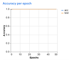
Training accuracy graph over epochs, showing model convergence approaching 99.51%.
Loss
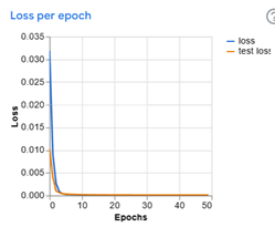
Training loss graph demonstrating steady decrease during model optimization.
Confusion Matrix
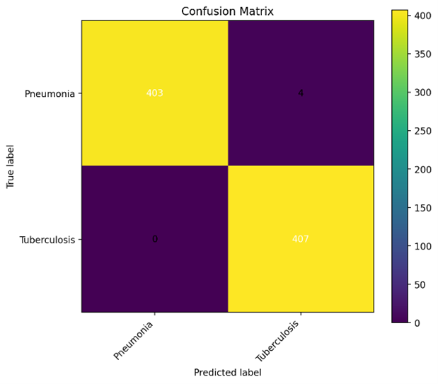
Matrix showing the accurate classification of Pneumonia and Tuberculosis cases with minimal false predictions.
Precision & Recall
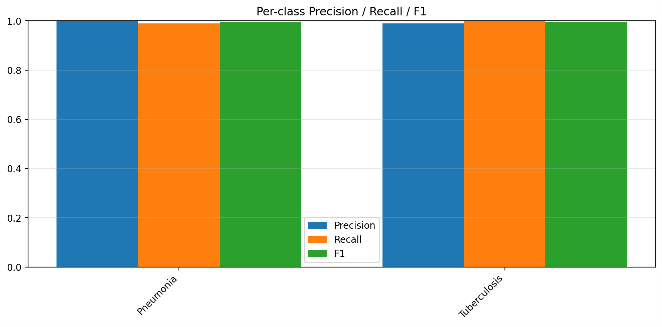
Evaluation showing high macro-precision (99.51%) and macro-recall (99.51%), with macro-F1 score of 99.51%.
ROC Curve
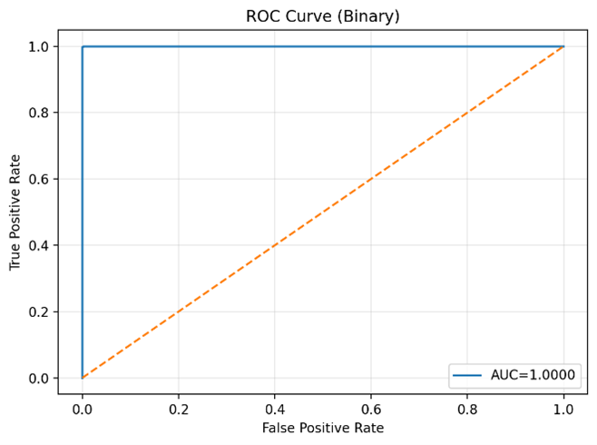
Receiver Operating Characteristic curve indicating excellent discriminative performance with ROC-AUC of 0.999994.
Research Methodology
Our approach to developing this AI diagnostic tool
Data Collection
We collected over 3,000 labeled chest X-ray images from multiple public datasets, ensuring diverse representation of normal cases, pneumonia, and tuberculosis.
Data Preprocessing
Images were standardized, normalized, and augmented to increase model robustness, with careful preprocessing to preserve medical diagnostic features.
Model Training
Multiple deep learning architectures were evaluated, with convolutional neural networks showing the best performance for classifying radiographic patterns.
Validation & Testing
Models were validated using cross-validation techniques and tested on independent datasets to ensure generalizability and minimize overfitting.
Web Implementation
The best-performing model was exported and implemented in this web application using TensorFlow.js and Teachable Machine for accessible deployment.
Future Directions
Next steps in our research journey
Expanded Disease Coverage
Extending our model to detect additional lung conditions including lung cancer, COPD, and interstitial lung diseases.
Clinical Validation
Conducting rigorous clinical trials to validate our system's performance in real-world healthcare settings.
Mobile Application
Developing lightweight mobile applications for offline use in remote areas with limited internet connectivity.
Educational Integration
Creating training modules for medical students to learn radiographic pattern recognition with AI assistance.
Research Disclaimer
This system is developed for research purposes and is not FDA/CE approved for clinical use. The AI predictions should not replace professional medical diagnosis, but rather serve as a supplementary tool to assist healthcare providers. Always consult qualified medical professionals for proper diagnosis and treatment decisions.